📖 PI-RADS 2.1 Assessment Categories
PI-RADS 2.1 employs a 5-point scale based on the likelihood that mpMRI findings correlate with clinically significant cancer.
Clinically significant cancer is highly unlikely to be present. These lesions typically require no further follow-up.
Clinically significant cancer is unlikely. These findings generally do not warrant immediate biopsy in the absence of other risk factors.
The presence of clinically significant cancer is equivocal. Management should incorporate additional clinical factors such as PSA density.
Clinically significant cancer is likely. Biopsy is strongly recommended, with MRI-targeted approaches preferred.
Clinically significant cancer is highly likely. These lesions mandate tissue diagnosis and expedited clinical management.
Peripheral Zone – DWI Dominant
Peripheral Zone Scoring Algorithm:
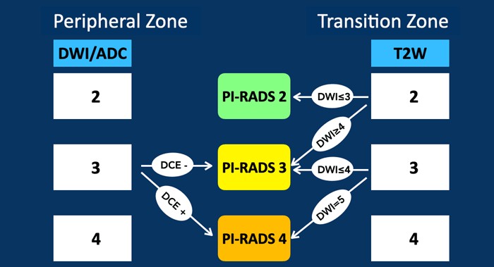
- DWI=1 or 2: Direct PI-RADS 1 or 2 (no DCE needed)
- DWI=3: Requires DCE: negative→PI-RADS 3; positive→PI-RADS 4
- DWI=4 or 5: Direct PI-RADS 4 or 5 (no DCE needed)
Transition Zone – T2W Dominant
Transition Zone Scoring Algorithm:
- T2W=1: PI-RADS 1 (no DWI needed)
- T2W=2: DWI ≤3 → PI-RADS 2; DWI ≥4 → PI-RADS 3 (upgrade)
- T2W=3: DWI ≤4 → PI-RADS 3; DWI=5 → PI-RADS 4 (upgrade)
- T2W=4 or 5: Direct PI-RADS 4 or 5 (no DWI needed)
PI-RADS 2.1 Assessment Tables
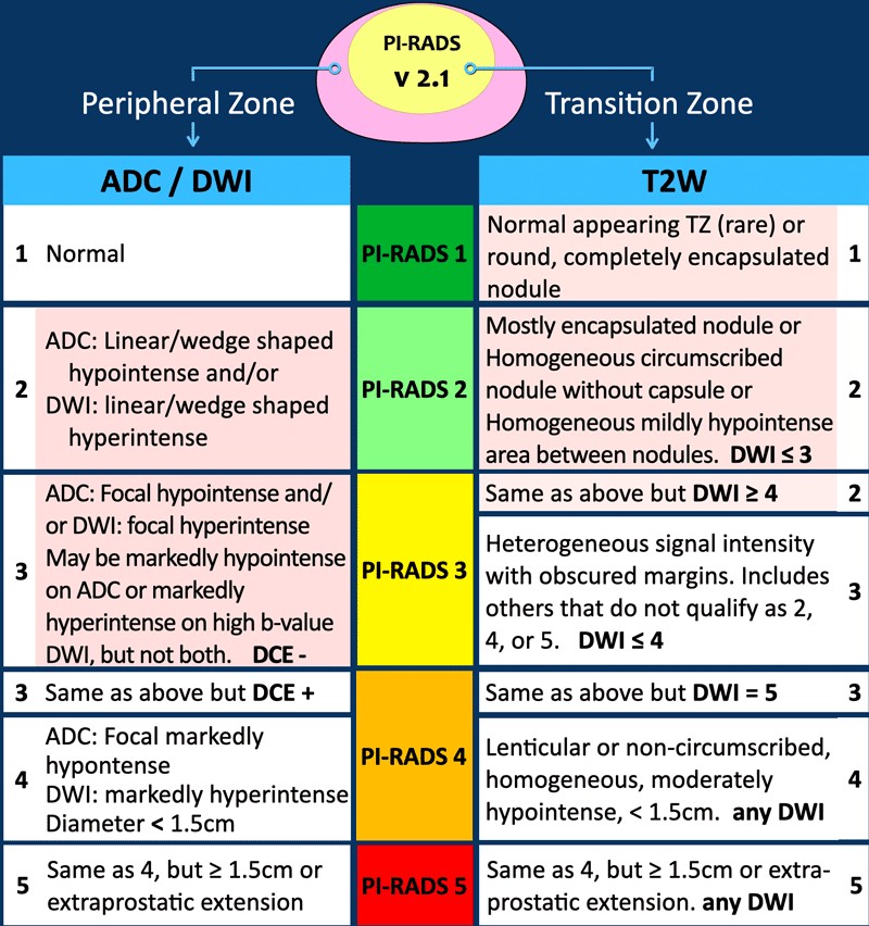
Official ACR/ESUR v2.1 cross-tables. In the peripheral zone, DWI is dominant and DCE is only a tie-breaker for DWI 3; in the transition zone, T2W is dominant and DWI upgrades the equivocal categories.
Peripheral Zone (DWI dominant)
| DWI | DCE | PI-RADS |
|---|---|---|
| 1 | Any | 1 |
| 2 | Any | 2 |
| 3 | Negative (−) | 3 |
| 3 | Positive (+) | 4 |
| 4 | Any | 4 |
| 5 | Any | 5 |
Transition Zone (T2W dominant)
| T2W | DWI | PI-RADS |
|---|---|---|
| 1 | Any | 1 |
| 2 | ≤ 3 | 2 |
| 2 | ≥ 4 | 3 2+1 upgrade |
| 3 | ≤ 4 | 3 |
| 3 | 5 | 4 3+1 upgrade |
| 4 | Any | 4 |
| 5 | Any | 5 |
Size / EPE rule: a lesion otherwise scored 4 is upgraded to PI-RADS 5 when it measures ≥ 1.5 cm or shows definite extraprostatic extension.
Whole-gland score: equals the highest-scoring (index) lesion.
PI-RADS v2.1 Decision Flowcharts
Visual summary of how the peripheral zone (ADC/DWI-dominant) and transition zone (T2W-dominant) scoring algorithms combine into the final PI-RADS assessment category.
Clinical Decision-Making
PI-RADS 1-2 (Low Suspicion): Biopsy not recommended absent other compelling factors. Negative predictive value is high.
PI-RADS 3 (Equivocal): Management varies by location and clinical factors. Consider PSA density, follow-up MRI, or risk-stratified approaches.
PI-RADS 4-5 (High Suspicion): Biopsy recommended. MRI-targeted approaches show superior detection compared to systematic biopsies alone.
Key Technical Parameters
- Field Strength: Both 1.5T and 3.0T acceptable; 3.0T preferred for superior SNR
- DWI b-values: ADC with b=0-100 and b=800-1000 s/mm²; high b-value DWI at b=1400-2000 s/mm²
- DCE Temporal Resolution: <15 seconds to capture early enhancement
- Post-biopsy Interval: ≥6 weeks for staging purposes
Version 2.1 Updates from 2.0
Version 2.1 clarified several ambiguities from version 2.0:
- Improved transition zone scoring with explicit upgrading rules (2+1 and 3+1)
- More precise DWI criteria distinguishing focal from linear/wedge-shaped abnormalities
- Clarified measurement standardization and prostate volume calculation
- Enhanced peripheral and transition zone morphologic descriptions
Diagnostic Performance
Clinically Significant Cancer Detection (PI-RADS ≥4):
- Sensitivity: 0.83-0.90
- Specificity: 0.48-0.66
- Area Under Curve: 0.86-0.90
Inter-reader Agreement (Kappa): 0.40-0.60 (moderate to good). PI-RADS 3 has greatest variability.
Prostate Cancer TNM Staging
Standard AJCC/UICC TNM staging for prostate cancer (8th edition). MRI findings such as extraprostatic extension (T3a) and seminal vesicle invasion (T3b) directly inform clinical T-stage.
| Category | Definition |
|---|---|
| T — Primary Tumor | |
| T1 | Clinically inapparent tumor, not palpable or visible by imaging |
| T2 | Tumor confined within the prostate (T2a ≤ half of one lobe; T2b > half of one lobe; T2c both lobes) |
| T3a | Extraprostatic extension (unilateral or bilateral), or microscopic bladder neck invasion |
| T3b | Tumor invades seminal vesicle(s) |
| T4 | Tumor is fixed or invades adjacent structures other than seminal vesicles: external sphincter, rectum, bladder, levator muscles and/or pelvic wall |
| N — Regional Lymph Nodes | |
| N0 | No regional lymph node metastasis |
| N1 | Regional lymph node metastasis |
| M — Distant Metastasis | |
| M0 | No distant metastasis |
| M1a | Non-regional lymph node(s) |
| M1b | Bone(s) |
| M1c | Other site(s), with or without bone disease |
References
- American College of Radiology. PI-RADS Prostate Imaging – Reporting and Data System, Version 2.1. ACR, 2019.
- Turkbey B, Rosenkrantz AB, Haider MA, et al. Prostate Imaging Reporting and Data System Version 2.1: 2019 Update. Eur Urol. 2019;76(3):340-351.
- Weinreb JC, Barentsz JO, Choyke PL, et al. PI-RADS Prostate Imaging – Reporting and Data System: 2015, Version 2. Eur Urol. 2016;69(1):16-40.
- The Radiology Assistant. Prostate Cancer – PI-RADS v2.1. radiologyassistant.nl.
- Amin MB, Edge SB, et al. (eds). AJCC Cancer Staging Manual, 8th ed. — Prostate. American Joint Committee on Cancer, 2017.
Medically reviewed by Dr. Amar Udare, MD, DNB · Last reviewed: